
Koshits Ivan Nikolaevich
Petercom-Network Management System/Consulting Grope Close Corporation, Russia
Title: Optical features of the anterior segment of the eye and lens
Biography
Biography: Koshits Ivan Nikolaevich
Abstract
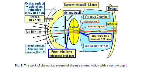
1. Optics of the anterior segment of the eye. We are used to idea that light rays entering the eye are refracted by the cornea. However, this is not the entirely true. Incoming light rays first pass through the first barrier: the tear film with the epithelium, where the refractive index (RI) = + 43.6 D on the border with air. And only then the rays pass through the second barrier "tear film – cornea" with RI = + 5.3 D. There is no air layer between the epithelium and the cornea, which makes it possible to create a kind of biological optical system: "tear film + epithelium + cornea". Such optical design of anterior segment of the eye produces a kind of" optical compression", it "collects" a wide picture of the visual space in the eye in a narrow" optical tunnel", as in a telescope. This optical compression is necessary in order to provide the possibility of passing the light bundle even through a narrow pupil (Pic. 1).
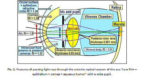
The thickness of the cornea in the center is almost 2 times less than in the periphery, which is typical for the scattering lens. Therefore, for example, the white light rays coming out of the cornea at the border of the "cornea – intraocular fluid" will be slightly divergent (see Fig. 1) and further subject to dispersion, decomposing into a spectrum, this means that inside the eye there will always be rays of all colors of the spectrum and only in the center of the cornea wavelengths of light will all be in one bundle – the white light. Furthermore, the anterior optical system (AOS) of the eye, consisting of the "eye surface" and the cornea, borders with a less dense optical medium – intraocular fluid. AOS works as a biological lens, providing at first compression in the "light tunnel", and then - a weak scattering of passing light rays.
Therefore, white light rays will always be subject to dispersion on the posterior surface of the cornea, and will go to the anterior surface of the lens as slightly divergent and expanding color bands, including three base bands: blue, green and red (BGR-band), which can be detected by macular cones. The effect of thickness and topography of the tear film with corneal epithelium on the overall refractive power of the eye is quite noticeable. Therefore, in dry eye syndrome, patients often complain about the lack of image sharpness, since the thinning of the lacrimal surface significantly reduces its refractive power.
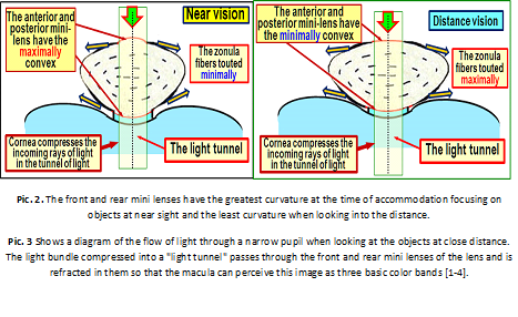
2. Optical structures of the lens. The mystery of the focusing mechanism of light rays that pass close to the optical axis of the eye exactly into the macula for many years had no solution. Traditionally, it was believed that the lens capsule cannot strongly refract light in its central region due to the small curvature radius. As it turned out, this is not a complete picture. In the center of the anterior and posterior surfaces of the lens capsule there are two thin mini-lenses: the front mini-lens and the rear, which change their optical powers depending on the pressure inside the lenses capsule, depending on the tone of the ciliary muscle (Pic. 1). These mini lenses provide maximum refraction when looking at the objects at the minimum distance. Pic. 2 shows the change in the geometry of the front and rear mini-lens in the extreme phases of accommodation. When looking at closely located objects the pressure in the capsule of the lens is as big as its refractive power (the circumference) of these mini-lenses. When looking into the distance, the pressure inside the lens bag same as the refractive power of the mini lenses are at its minimum. This is because when you look objects placed at near distance the capsule of the lens is the least stretched by the ciliary zonules and therefore the most powerfully compresses structure inside the lens.
Conclusion. The anterior optical segment of the eye is a composite biological lens, which includes the following refractive structures: tear film, corneal epithelium and the cornea itself. The main task of the eyes optic system is to effectively compress into a "light tunnel" with a diameter of 1.8 -2.5 mm coming into the eye light flux for it to pass even through the narrow pupil. The cornea is a weakly scattering lens, so the light rays are spread out in the spectrum at the border with the aqueous humor of the anterior chamber, then undergo a weak deviation from the outside on the posterior surface of the cornea. In the center of the optical axis in the lens capsule there are two mini-lenses – anterior and posterior, which change their curvature (refractive power) in different phases of accommodation due to changes in pressure inside the lens with different tone of the ciliary muscle.